FTIR Microscope Thermo Fisher Nicolet iN10
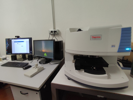
FTIR Thermo Fisher Nicolet iN10 Microscope for the acquistition of high spatially resolved Fourier Transform Infrared spectra (possibility to obtain infrared map with spatial resolution lower than 10 μm), in Transmission, Reflection and ATR measurment configurations.
Technical characteristics
Ever-Glo air-cooled infrared source
Microscope integrated Interferometer with minimum spectral resolution of 1 cm-1
Maximum spectral range available of 7800-350 cm-1 for the different detectors
Motorized stage XYZ, 70x127 mm
LED illuminantion separated for transmission and reflection configurations
Visible Light Polarizer for micro-plastic analysis
Supplies:
Slide-On MicroTip ATR system with a germanium crystal of 0,35mm Ø, for point ATR measurements and mapping in tapping-mode configuration
ATR-Imaging system with a germanium crystal of 1mm Ø, for ATR mapping of large sample portions through a singol compression (useful for soft samples measurements or to avoid the cross-contamination problem)
Micro-compression cell with diamond-anvil windows, for micro-transmission analysis of samples to be thinned (single fiber, mineral and crystal or paint chips)
Objective:
Cassegrain IR/VIS objective, permanently aligned 15X/0,7 FWD 16 mm
Detector
DTGS operating at room temperature (spectral range 7600-350 cm-1)
MCT-A liquid nitrogen cooled (spectral range 7800-650 cm-1)
Software
Complete Omnic software for microscope management, acquisition of image and spectra, mapping and data analysis. This sortware allows, trough Picta and Specta package, to performe the multivariate (MCP) and the principal component (PCA) analysis for IR map. Moreover, allows the mupltispectral analysisi of single IR spectrum through the comparison with 40000 spectra.