History of the Center
In 1985 the Academic Senate determined the establiscment of the Electron Microscopy Center.
The decision was made right after the deal between some professors of tree Scientific Faculties (Science MM FF NN, Engineering, Medicine) with the aim of building a service structure for research, education and territory development, centred on microscopic characterization. This deal immidiately found support among many departments.
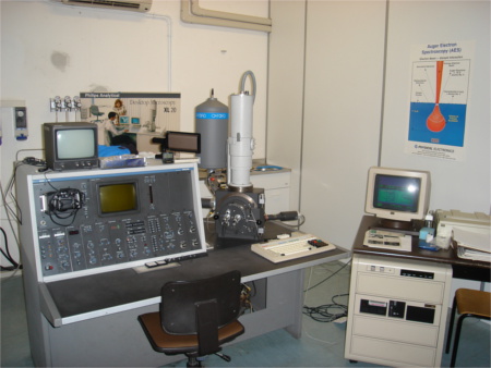
The start action consisted to buying a Scanning Electron Microscope (Philips SEM505, transferred to the University's Museaum since 2018), which has been installed at the Engineering Faculty in roio's campus. Over the following years, the instrumental equipment grew up, due to the University's investment policy. Tre preparative section for both DEM and Metallograohic was completed, the X-ray microanalysis for SEM got installed and an image analysis system was purchased. In these years, the Medical Faculty contributes to the development of the Center with the purchase of a TEM, installed to its laboratories.
In 1995, with the purchase of a Confocal Microscope (one of the first in Italy), was opened a new section at the Science Faculty in Coppito's campus. At this point, while maintaining a single management, the Center assumed a bipolar structure, articulating in two areas: physical-technological (at Roio's campus) and biomedical (at Coppito's campus).
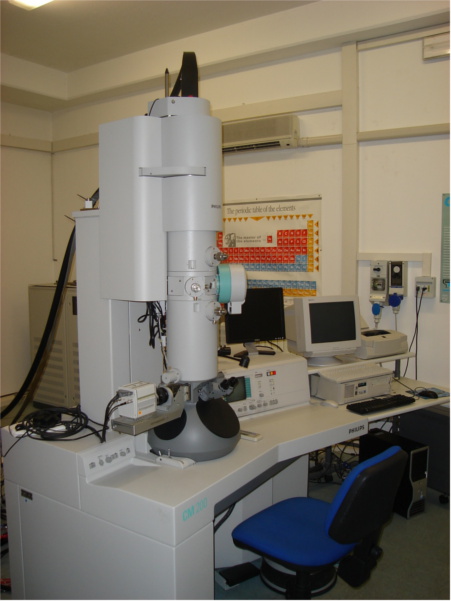
In the following years, a significant MIUR financing for large equipment, allowed the purchase of a new SEM that operates under controlled pressure, a TEM of 100kV for biological applications and a TEM of 200kV, equipped with microanalysis, for material science applications. Beyond that, a research optical microscope with an image analysis program has been purchased, and then a whole line of tools for the TEM preparation, for both biology and materials.
At that time, Engineering Faculty and some departments od Science Faculty provided ample spaces for the new equipmnet installation.
Thanks to the Center development in different microscopy field, tha name was changed from Electron Microscopy Center to Microscopy Center.
The tragic earthquake of 2009 stop the Center development, that start again this activity with grate difficulty in Coppito's campus. Indeed, the roio's campus was seriously damaged and will be closed for many years. In 2016, thanks to the approval of the new regulation with represntant of all University's department in the Board of Director, the Coppito's campus become definitely the only headquarter of the Microscopy Center.
In 2017, an important University's funding of "Grandi Progetti di Investimento" allow to build a new top class SEM (Zeiss GeminiSEM 500, equipped with differen detectors and additional features), that will allow to grow up the Center offer quality, both in terms of internal research activity than for the ervices for other research bodies and local companies.
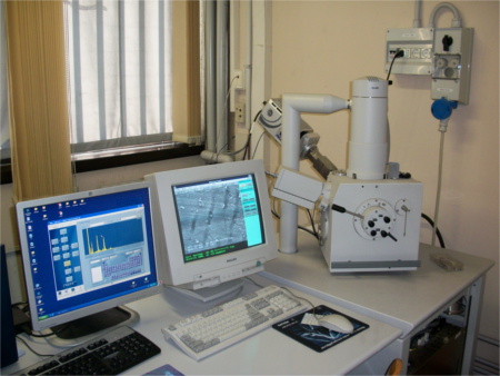
In 2018, beyond GeminiSEM installation, is made available to Center activity a FTIR spectrometer (Bruker Vertex 70v) for Infrared Spectroscopy measurements. In addition, the optical microscope Zeiss AxioImager is gratly improved to allow the Correlative Micrscopy between optical and scanning electron microscopies.
In 2019, a new FTIR microscope Thermo Fisher Nicolet In10 (equipped with ATR and supplied with a 30000 spectra's database), and a new camera with CMOS technology for the TEM CM100, are installed. Moreover, important upgrades are made on the Confocal Microscope (a new motorized stage and a new workstation) and on the Fluorescence Optical Microscope.
In 2021, an innovative zoom microscope Zeiss Axio Zoom V16, with a single optical path for the stereo acquisitions of reflected light, bright field transmitted light and fluorescence with optical sectioning, was bought.
The latest microscope purchased, in 2023, is an inverted microscope Zeiss Axio Observer for metallography. This inverted microscope, for acquisition on reflected light, combine different contrast method, from calssical to innovative ones, like dark and bright field, differential interference dontrast (DIC), circular polarized DIC (C-DIC), and polarization contrast.
The Directors
Prof. Pietro Picozzi, 1985-1993
Prof. Sandro Santucci, 1994-1997
Prof. Pasquale Carelli, 1998-2003
Prof. Guido Macchiarelli, 2004-2016
Prof. Luca Lozzi, 2017-2022
Prof. Maria Grazia Palmerini, from 2022
The technical staff
Mrs. Luisa Corona, 1985-1999
Dr. Maria Giammatteo, from 1985
Dr. Lorenzo Arrizza, from 2001
Dr. Angelo Sarra, from 2020